
Teaching mushroom microscopy is one of the most rewarding skills you can pass on, and also one of the most mishandled. Most guides focus on what to look at under the lens, not how to actually teach someone else to do it. That gap matters. Whether you are leading a university practical, running a mycology club session, or mentoring hobbyists one to one, knowing how to teach mushroom microscopy effectively changes outcomes. This guide covers the equipment, preparation workflows, teaching techniques, troubleshooting strategies, and assessment methods you need to deliver sessions that genuinely build skill.
Table of Contents
- Key takeaways
- How to teach mushroom microscopy: tools and preparation
- Preparing slides: a step-by-step teaching workflow
- Microscopy techniques: focusing, lighting, and magnification
- Common teaching challenges and how to address them
- Assessing learner progress in mushroom microscopy
- My honest experience teaching mushroom microscopy
- Equipping your sessions with Sporebuddies
- FAQ
Key takeaways
| Point | Details |
|---|---|
| Start with the right tools | Calibrated eyepiece micrometers and appropriate mounting media are non-negotiable for accurate teaching. |
| Slide prep beats magnification | Lighting and slide quality determine clarity far more than pushing to higher magnification. |
| Build skills incrementally | Begin learners on simple, common samples before advancing to complex or unfamiliar fungi. |
| Combine hands-on with visual aids | Pairing live observation with reference images significantly reinforces identification skills. |
| Assess with practical tasks | Structured observation goals and documented measurements give learners clear, measurable progress. |
How to teach mushroom microscopy: tools and preparation
Before you lead a single session, you need to be confident about the equipment in the room and the concepts you are introducing. An intro to mushroom microscopy works best when learners understand why each tool exists, not just how to use it.
For microscopes, a compound light microscope with objectives at 4x, 10x, 40x, and 100x (oil immersion) covers the full range of fungal microscopy work. Most routine identification tasks happen at 400x total magnification. Resolving hyphal features increases clearly between 400x and 1000x, so access to at least one oil immersion setup in a group session is ideal.
Calibrated measurement tools deserve special attention. Spore size is a primary identification feature; for example, Psilocybe cubensis spores measure approximately 11–17 x 7–12 μm, and a stage or eyepiece micrometre is the only reliable way to record those values accurately. Teach your learners to calibrate before every session, not once and never again.
Here is a summary of the core materials for any teaching session:
| Item | Purpose | Notes |
|---|---|---|
| Compound microscope | Primary observation tool | Objectives: 4x, 10x, 40x, 100x |
| Eyepiece micrometre | Spore measurement | Must be calibrated per objective |
| Spore prints | Primary sample source | Collect 12–24 hours before session |
| Microscope slides and coverslips | Slide preparation | Keep dust-free before use |
| Mounting media (water, lactic acid, glycerol) | Clarity and preservation | Each has different drying rates |
| Forceps and dissection needles | Hyphal material handling | Fine tips recommended |
| Reference images or field guides | Comparative identification | Printed or digital both work |
Pro Tip: Before teaching session day, prepare a small set of pre-made demonstration slides. These give learners a clear reference point and let you focus on explaining what to look for rather than troubleshooting everyone’s slide at once.
The beginner guide to mycology microscopy from Sporebuddies covers these fundamentals in detail and works well as a pre-session reading resource for learners.
Preparing slides: a step-by-step teaching workflow
Slide preparation is where most learners struggle first. Teaching a consistent, repeatable workflow removes a lot of that uncertainty. The spore print to slide workflow is one of the clearest ways to structure this for students.
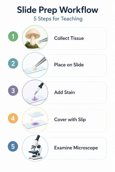
Follow this sequence when demonstrating slide preparation:
- Collect the spore print at least 12 hours before your session. A fresh print left overnight on white paper gives a visible deposit and confirms spore viability.
- Choose your mounting medium. Water is simple and good for quick observations. Lactic acid and glycerol are better choices for teaching because they dry more slowly, giving learners more time to adjust and reposition the coverslip without the image degrading.
- Place one drop of medium on a clean slide. Use a dissection needle to transfer a tiny amount of spore material from the print into the drop. Less is more. Overcrowded slides are one of the most common errors beginners make.
- Lower the coverslip slowly at an angle from one edge to avoid trapping air bubbles. This step benefits enormously from a live demonstration before learners try it.
- Warm the slide at around 50°C for five minutes. Slide warming at this temperature softens tissue and improves viewing clarity, especially for spores that have not fully separated.
- Blot any excess medium from the slide edges before placing it on the stage.
For hyphal preparations, cut a very thin section from gill tissue or mycelium using a sharp dissection needle and mount it in the same way. Teach learners to tease the material apart in the mounting medium rather than pressing down with the coverslip, which crushes fragile hyphal structures.
Pro Tip: Prepare one intentionally poor slide alongside a good one when demonstrating. Showing learners exactly what an overcrowded or bubble-filled slide looks like helps them self-diagnose their own errors quickly, rather than assuming every imperfect image is normal.
Microscopy techniques: focusing, lighting, and magnification
Once slides are prepared, the real teaching work begins. This is where the most persistent misconceptions about fungal microscopy methods surface, and where your guidance has the most impact.
The single most important habit to build in learners is starting at low magnification. Begin at 4x to locate the sample area, then move to 10x before committing to 40x. Many students jump straight to high power and then spend ten minutes unable to find anything. Starting low prevents that frustration entirely.
Lighting deserves as much attention as magnification. Correct lighting and contrast matter more for fungal identification than reaching maximum magnification. Teach learners to lower the condenser and reduce light intensity when first scanning at low power, then gradually increase illumination as they move to higher objectives. Lowering light for initial detection, then raising it progressively, consistently produces better results than leaving brightness at a fixed setting throughout.
Key structures to teach learners to identify:
- Spore shape and size. Terms to introduce: ellipsoid (oval), subglobose (nearly spherical), fusiform (tapered at both ends). Each has a clear visual analogy that helps novices.
- Spore surface ornamentation. Smooth, verrucose (warty), or striate surfaces are visible at 400x with good slide preparation.
- Septation in hyphae. Cross walls (septa) separating hyphal compartments are clearly visible at 400x in many species.
- Clamp connections. These small, bracket-like structures at hyphal septa are a key identification feature in many basidiomycetes. Teach learners to look for them specifically rather than discovering them by accident.
The mushroom spore shapes guide on the Sporebuddies site is a practical reference for introducing spore morphology vocabulary to learners.
Pro Tip: Keep a printed or digital reference sheet of spore morphology images at each workstation. Comparing live observations with reference material reinforces identification skills far more effectively than asking learners to rely on memory alone.
Common teaching challenges and how to address them
Even well-prepared sessions run into difficulties. Knowing the likely stumbling blocks lets you address them before they erode learner confidence.
“Microscopy skills develop gradually. The goal is not perfection in a single session but building a reliable process that improves with every slide.”
The most frequent issues educators encounter include:
- Difficulty focusing at higher magnification. This usually means the slide is too thick or the initial focus at low power was not precise enough. Teach learners to focus carefully at 10x before moving up, and remind them that the focus changes only slightly between 10x and 40x on a parfocal microscope.
- Low contrast and unclear images. This almost always reflects a problem with lighting or slide preparation rather than the microscope itself. Check condenser position and light intensity first.
- Overcrowded slides. A common beginner error. A slide with too much material produces a chaotic image where nothing is identifiable. Reinforce the habit of using minimal sample material from the first session onwards.
- Misconceptions about magnification. Many learners believe that higher magnification automatically means better identification. Address this directly. Slide preparation and lighting are the determining factors for image quality, not the objective used.
Microscopy skills build through repetition and graduated exposure to different sample types. Start learners on common, structurally simple species before introducing specimens with finer or more complex features. Confidence built on reliable early successes carries forward into more challenging work.
On the topic of molecular data: it is worth explaining to learners that microscopy remains a rapid and cost-effective first step in identification, complementing rather than competing with DNA sequencing. Framing it this way prevents learners from dismissing microscopic observation as outdated.
Assessing learner progress in mushroom microscopy
Good assessment in mushroom science education does not have to be complex. What it does need to be is consistent and tied to observable outcomes rather than vague impressions of how a learner is doing.
Use this framework to structure your assessment:
- Set specific observation goals. For example: “Identify and sketch the spore shape, measure the long axis, and note surface ornamentation for three different samples.” These are verifiable and give learners a clear target.
- Use practical exams with prepared samples. Provide a set of slides and ask learners to document what they observe. This mirrors real mycology work and tests both technical and observational skills together.
- Require measurement records. Ask learners to record spore dimensions in every session. Over time, these records show whether measurement accuracy and consistency are improving.
- Encourage participation in citizen science or local mycology groups. Real-world field work and community projects accelerate skill development in ways that classroom sessions alone cannot. The UK mushroom identification guide from Sporebuddies works well as a field reference for learners moving from lab to field.
- Provide written feedback with specific next steps. Vague encouragement does not build skill. Telling a learner exactly what to practise next session does.
My honest experience teaching mushroom microscopy
I have watched learners spend an entire session chasing the highest magnification objective, convinced that resolution solves everything. It rarely does. What I have consistently found is that the educators who get the best results are the ones who spend the first thirty minutes of any session solely on lighting and slide preparation before a single observation is recorded. The technical side of using a microscope is learnable in an afternoon. The judgement to prepare a clean slide, set up lighting correctly, and read what you are seeing is built over many sessions.
The other thing I keep returning to is this: patience with beginners pays off disproportionately. Learners who are given simple samples early and allowed to succeed before complexity increases tend to develop genuine curiosity. They start asking questions you have not anticipated. That curiosity is harder to teach than slide technique, and it is far more valuable in the long run.
I also think the teaching community underestimates how much good reference material changes outcomes. Combining live observation with printed or digital comparison images is not a shortcut. It is how real mycologists work, and it is exactly what you should be modelling in your sessions.
— Fabio
Equipping your sessions with Sporebuddies
If you are building or expanding a mushroom microscopy teaching programme, having reliable, quality-sourced materials makes a genuine difference to what your learners can achieve. Sporebuddies supplies a full range of mycology equipment and supplies suited to both academic settings and enthusiast groups, including microscopes, slides, and mounting essentials.
For sample material, the mushroom spore syringe range offers clearly documented strains well suited to microscopy study. Institutions and group organisers running regular sessions can also explore the wholesale spore options to supply multiple learners cost-effectively. The mycology science and education section of the site brings together guides, resources, and product recommendations specifically aimed at educators and researchers who want to teach this subject well.
FAQ
What microscope magnification is best for mushroom microscopy?
Most fungal identification work takes place at 400x total magnification. Oil immersion at 1000x is used for finer hyphal details, but correct lighting and slide preparation matter more than reaching maximum magnification.
How do you prepare a spore print slide for teaching?
Collect a spore print 12 to 24 hours before the session, transfer a small amount of material into a drop of lactic acid or glycerol on a clean slide, lower the coverslip slowly, and warm the slide at around 50°C for five minutes before observing.
What are the most common errors beginners make in mushroom microscopy?
The most frequent mistakes are overcrowding slides with too much material, jumping directly to high magnification without focusing at low power first, and neglecting to adjust lighting and condenser position.
How long does it take to teach competent mushroom microscopy skills?
Building reliable observational and identification skills typically requires multiple sessions over several weeks, starting with simple samples and progressing gradually. Consistent practice and access to reference material are the key factors.
Can mushroom microscopy be taught effectively to hobbyists as well as students?
Yes. The core workflow of slide preparation, lighting adjustment, and structured observation translates directly to hobbyist settings. Starting with well-documented species and clear reference guides keeps the learning curve accessible.